
What are You Made Of?
Think
Do you ever think about what you are made of and how your body works to keep you breathing, thinking, and moving? Think about all of your body parts, and how they work together. In the box, describe in as much detail as you can, how your body performs an action like running, or catching, or writing.
When you catch a ball, lots of parts work together. You see the ball with your eyes, your brain guesses where it’s going to go, and it sends a message to your body (arms and hands) to catch it. If you go deeper, cells in your eyes receive a signal and send it to your brain. Your brain sends a signal to the cells in your arms, that work together and move you. In this section, we will look at how these cells work together.
In this learning activity we will learn about the smallest unit of life, the cell, and how different cells in our body look, replicate, and work together. We will start by reviewing the different parts of the cell and learn how the cell replicates or divides.
But before we begin we should discuss the culminating activity for this course which is worth 15% of your final grade and is something that you will be thinking about and working on throughout the course. In this culminating activity, you will explore how what you have learned in the course impacts a career of your choice.


Your task is to research a career that you find interesting and discover how science plays a role in that career. This project has 5 parts and you should be working on each part as you work through the course. Each part will be a one- page visual summary/poster and should contain images and written content. Make sure you use headings to organize your thoughts and include images. For more information about the culminating activity, visit Learning Activity 4.7 Researching Careers. One more thing, during this course you will take three quizzes each worth 5%. Be sure to try the practice quiz before you do the actual quiz for grades.
Setting up a Notebook
While working through this course, you will need one other tool to help you keep organized. You will need a “notebook”. This may be an actual pen and paper notebook where you can take notes and write reflections as instructed throughout the course. Or, you may choose to have a digital “notebook” or a file where you type in notes and reflections. This is a personal decision, but it is important that you do have a place for notes. Be sure to add a title to each set of rough notes with the learning activity title and number. Jotting down words and scientific terms in your notebook is an effective way of keeping track of the new vocabulary that you will be learning throughout the course.
You may use a number of different methods for taking journal entries. These may include:
- Cornell note-taking system
- T-charts
- Visual notetaking (sketchnoting)
- Mindmaps
Who Discovered the Cell and How Do We Know all of This?
In 1665, Robert Hooke (the legendary London architect, inventor, and scientist) was looking at a cork sample under his latest invention, the microscope. Cork is an important tissue found in plants, and it is the same cork you find sealing wine bottles. The tiny structures that he observed reminded him of the small chambers in monasteries in which monks (holy men) lived. Each narrow bed chamber was called a “cell.” Hooke decided then that these small structures could also be appropriately named cells.
The Cell is the Basic Unit of Life
The discovery of the cell led to a flurry of research and curiosity about cells and how they relate to all living things. For instance, microscopes revealed that there were cells in the skin of an onion, as well as in the skin of a person. Since then, a lot has been researched and discovered about cells; what they are, how they function, and how we can apply this knowledge when cells become diseased.
It is now widely known by scientists that the cell is the basic unit of life. In fact, the basic importance of cells and how they function is summarized in an explanation with three parts, called the Cell Theory.
“Theory” is a very important word in science. It DOES NOT mean the same thing as “Idea” or “Best Guess”. “Theory” is a word that scientists use instead of saying, “a well-proven explanation of how something works.”
Cell Theory
The foundation of what we know about growth and reproduction is summarized by the cell theory. Because it has not been disproven, this theory is widely accepted by cell biologists. For centuries, the three statements of cell theory have been the basis for research conducted on cells and diseased cells, such as cancer cells.
Here are the three statements of cell theory:
What’s inside a cell? Let’s take a Cell-fie!

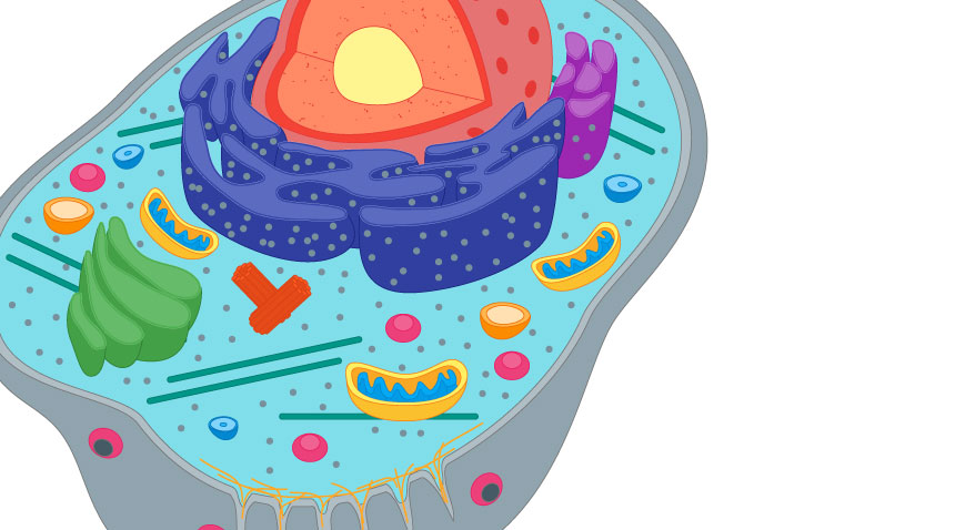
Before we get into how and why cells divide, we need to have an understanding of the different parts and what they do. Later in the learning activity, you will see how different cells in the body have the same parts, but use them differently.
The different parts of the cell are called organelles. The word organelle means, “little organs”, and just as you know that different organs in your body have different jobs, different organelles in a cell have different jobs.
Parts of the Animal Cell
All living things are composed of one or more cells. Cells have structures that enable them to ingest food, excrete waste, and in some cases, move around.
Watch This!
Watch this short video as an introduction to the structures of cells. (Opens in new window)
You may want to watch more than once. The resource link has been provided as a suggestion only. You are encouraged to conduct your own research to support your learning.
You can explore the different parts of the cell in any of these interactives.
Eukaryotic Organelle Cell Model (Opens in new window)
Cell explorer – The Animal Cell (Opens in new window)
Cell game (Opens in new window)
You may want to watch more than once. The resource link has been provided as a suggestion only. You are encouraged to conduct your own research to support your learning.
Create and complete a table similar to the one below using information from the interactives above.
| Organelle | Function |
|---|---|
|
Cell Membrane |
Is the outer boundary of the cell. It is semipermeable meaning it lets some things pass across and blocks other things from entering or exiting the cell. |
|
Nucleus |
Controls all activities in the cell and contains DNA. It is found in the cytoplasm. |
|
Ribosomes |
Make proteins. Found in the cytoplasm and also the rER |
|
Endoplasmic reticulum (ER) |
Rough ER (rER) has ribosomes, and smooth ER does not; it is located near/around the nucleus Transports proteins, food, water, and waste |
|
Mitochondria |
Sometimes called the “Power house” of the cell. Makes energy for all the cells activities |
|
Golgi apparatus |
Packages proteins |
|
Centrioles |
Help cells divide |
|
Cytoplasm |
Jelly-like substance that suspends the organelles, holding them in place while allowing some movement. |
|
Vacuole |
Stores water, food, and waste |
Let’s see if you can do some Animal Cell Labelling based on what you remember.

A Closer Look at Cells - Microscopy
Since its invention, the microscope has made tremendous contributions to the progress of science. Most of what we know about normal cells and diseased cells is due to the development of the microscope. Often, the word “microscope” refers to the standard classroom “compound light microscope,” but there are other types. Here, you’ll learn a little more about the two main types.
Light microscope:
A light microscope uses light (rather than electrons) to view a small object, such as a cell specimen. This compound microscope uses two lenses to magnify an object up to 2000 times (2000x). Light from above or beneath is used to illuminate objects. These microscopes are useful for viewing full-colour images of living or non-living cells and tissues, but not for viewing tiny virus particles, molecules, or small organelles. Take a look at the compound microscope in a little more detail, in the following labelled diagram.

Electron microscope:
These microscopes are much more powerful than light microscopes. There are several different types that can magnify a specimen up to two million times. Transmission electron microscopes produce black and white images of cells that are no longer living. Before a specimen can be viewed, it is freeze-dried, coated with metal or graphite, placed in a vacuum chamber, and bombarded by electrons. Images are recorded digitally and displayed on a computer screen or printed on paper. A scanning electron microscope uses electrons and scans the outer surface of the specimen with them. A 3-D image is formed and the specimen can be living or non-living.

Light microscopes are used to view cells and tissues, while electron microscopes are used to view molecules and very tiny organelles. Let’s have a look at some images.


Using a Microscope
Microscopes need to be handled with care. When carrying a microscope, one hand should be on the microscope’s arm, and the other on the base. The microscope should be stored with the cord wrapped around the base, and the revolving nosepiece turned to low power.
Watch This!
Watch the short video to learn how to use a light microscope. (Opens in new window)
The resource link has been provided as a suggestion only. You are encouraged to conduct your own research about microscopes to support your learning.
Each part of the microscope has a specific function: You will explore the parts of the microscope by clicking on each part in this interactive animation.
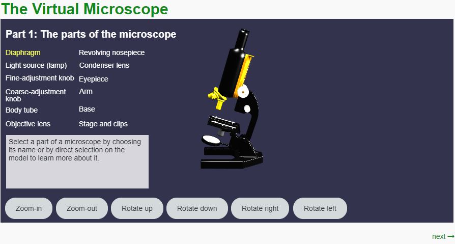
After completing the interactive, create and complete a table like the one below, listing the parts of the microscope and what they do.
| Part | Function |
|---|---|
|
Diaphragm |
|
|
Light Source |
|
|
Fine Adjustment Knob |
|
|
Coarse-adjustment knob |
|
|
Body tube |
|
|
Objective lens |
|
|
Revolving nosepiece |
|
|
Condenser Lens |
|
|
Eyepiece |
|
|
Arm |
|
|
Base |
|
|
Stage and Clips |
Magnifications of a Compound Light Microscope
As mentioned previously, you can revolve the nosepiece to choose different magnifications for viewing specimens. To “zoom in” on an object, you must switch to a higher magnification and then adjust the focus. The transition is sudden. It is as if one moment, you are standing on the other side of the room from a person and the next moment, you are 5 cm away from them. After zooming in, the object appears bigger and closer. You see less of the object, but more detail is revealed.
When determining the magnification of a specimen using a compound microscope, the magnification of the ocular lens is multiplied by the magnification of the objective lens. For example, if the ocular lens magnifies 10× and the objective lens magnifies 40×, the total magnification would be 10 × 40 or 400×.
This means that the specimen being magnified under the microscope appears 400× larger than the actual specimen. Try the following example.
Example:
If the magnification of the ocular lens is 5× and the objective lens is 25×, what is the total magnification?
Solution:
The total magnification would be 5 × 25 or 125×.
You will learn more about proper use of the microscope, in the next section.
Biological Drawings
Often, when you view objects under the microscope, you will also draw a proper biological diagram by hand, to make proper observations of the specimen being viewed.
Here are some tips for drawing proper biological diagrams by hand:
- Drawings include labels, titles and other information should be done with pencil.
- The title of the drawing is simply the name of the object you are looking at. For example: “Human red blood cell smear”
- Use unlined white paper.
- The drawing should be as large as possible (at least one-third of the page) and should be kept to the left of the centre of the page.
- All labels must be neatly printed and lined up on the right-hand side of the drawing. Use a ruler for label lines.
- Each cell is different. The drawing should be an outline of what you see. Do not include additional structures just because you think you should be seeing them.
- Do not shade or fill in areas with colour. All lines should be solid and complete.
Here’s an example of a scientific drawing. It is of human cheek cells.

Try It!
Identify the features of the images that make each one a good or bad drawing.

The top drawing is neat with smooth, deliberate lines, no sketching or colouring in, lines are done with a ruler and do not cross over each other; all labels are neat on the right hand side and written horizontally;
The bottom drawing is not a good biological drawing because the lines are messy, there is shading in of the structures, the lines cross over each other and the labels are not aligned.
Both diagrams require a label.
A Closer Look at Cells - How Cells Divide
Cell division is essential to multicellular organisms. Give some reasons why cells need to be divided and replicate themselves.
Cells divide to:
- Replace old, dead, or damaged cells
- Because they are getting too big
- Allow an organism to grow
- For reproduction
Living things are all capable of growth, but have you ever actually tried to picture HOW this happens?
Firstly, a cell which is formed with the combination of a female egg cell and a male sperm is a zygote. It’s hard to believe that you began as a single cell about the size of the dot in an exclamation point! How did you become the large mass of trillion cells that you are made up of today? Your first cell divided into two cells, called daughter cells. Those cells then divided into two more daughter cells, and so on, in a seemingly endless repetition of cycles.
View the video “Mitosis” to introduce yourself to the cell cycle.
As you learn the details of the cell cycle you will be invited to re-watch specific sections of this video in order to visualize what is happening inside the cell.
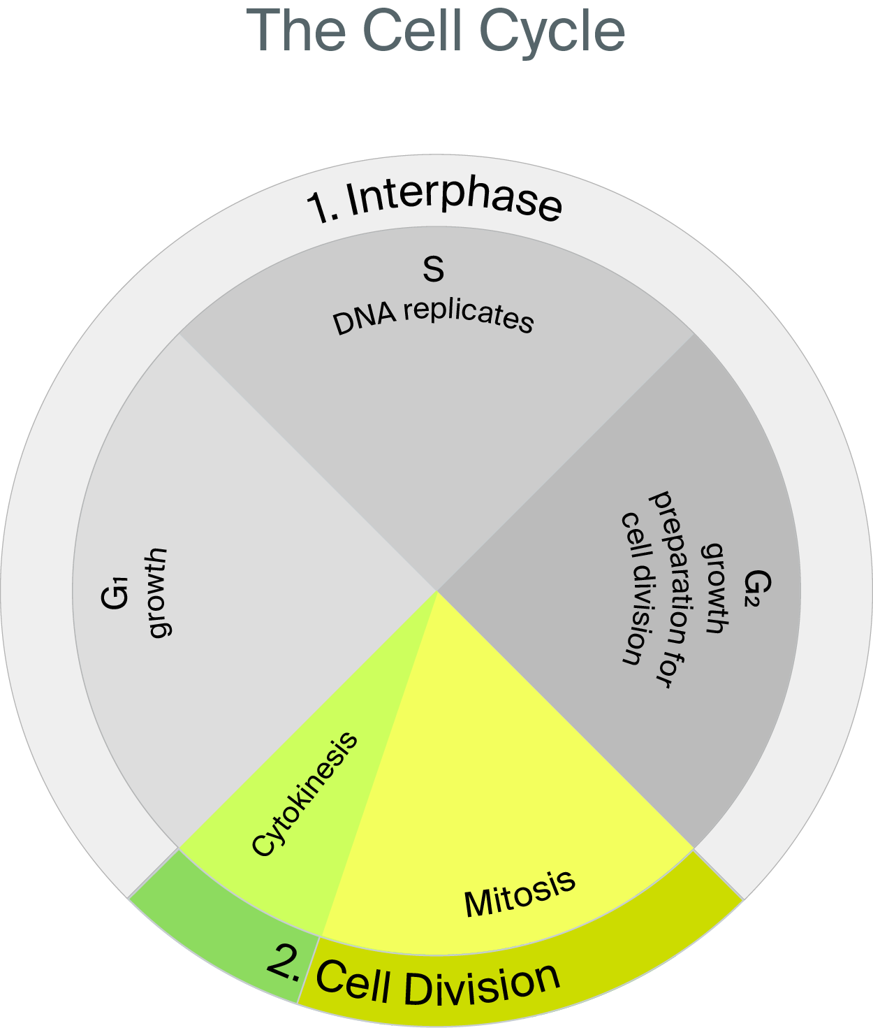
Observe the image below to summarize what you saw in the video. It is astonishing how just one single cell can divide and change into a fully developed person.
The cycle is a repeating sequence of events or activities. For instance, you could think of the four daily activities of an elderly cat as a cycle. Imagine a cat that spends 90% of its day sluggishly resting, then eating, and then resting again. It then spends 10% of its time actively playing and looking through windows. Similarly, most cells follow a process of slow growth and active division called the cell cycle.
Just as the house cat has periods of time during which it is sluggish or active, the cell cycle also has periods of time during which the cell is dividing, and periods during which the cell is growing and performing its normal functions. A good way to represent the cell cycle would be a wall clock or a pie chart. The following pie chart shows the sequence of the activities in the cell cycle that occur in an animal cell. Ninety percent of the cell cycle consists of a period of growth called Interphase (including G1, S, and G2 phases), while 10% consists of a period of cell division (mitosis and cytokinesis phases). This is probably a good time to begin using your notebook as you are about to be introduced to lots of new terms.
Two Distinct Stages of the Cell Cycle
The amount of time it takes to complete one full cell cycle can depend on a number of things. For instance, some types of bacteria cells can divide rapidly, performing the entire cycle every 20 minutes, while human cell cycles can take 8 to 24 hours. The cell cycle varies from species to species, but also from cell type to cell type. All cells in the human body do not divide at the same rate. The lifespan of a cell depends on the “wear and tear” of that cell, which is a result of its location.
Rapidly-reproducing cells are found in skin, hair, intestine linings, and stomach linings. These cells are in contact with the sun, bacteria, food, and the outside environment continually.
Cells that reproduce more slowly are found in organs such as the heart, eyes, and kidneys (among others).

The initial division of the cell cycle is into 2 parts: Interphase and Cell Division. A cell spends 90% of its life in the first period of the cell cycle, called interphase. Interphase can be further broken down into three distinct phases called first growth or gap 1 (G1), synthesis (S), and second growth or gap 2 (G2).
Interphase stage: Consists of 3 Distinct Phases
Cell Division: Mitosis and Cytokinesis
Recall that the cell-division period has two easily observable phases called mitosis and cytokinesis.
Before continuing to read consider all you have learned in this learning activity and state 3 reasons you think cells need to divide.
- Growth
- Repair (healing)
- Replace (to replace dying cells)
- Reproduction of single celled organism
It is the fate of many cells in your body, such as skin and red blood cells, to be continually replaced. Skin cells eventually dry out and flake off. A blood cell becomes less efficient over time and must be replaced every 100 days or so. This is part of the normal maintenance and repair of your skin and circulatory system and is the reason why the tissues that produce skin and blood cells are always dividing. Press each one to learn more:
Mitosis Phase
The first two phases of cell division are called mitosis. Watch the portion of the video below before reading the details below. You may come back to watch this again if you find it helpful to visualize the phases of mitosis.
Mitosis is the division of the nucleus into two nuclei. Although mitosis occurs seamlessly and without interruptions, scientists divide this phase into four main sub-phases. These observable sub-phases or stages are illustrated in four diagrams, to help you better understand the process. The four stages of mitosis are prophase, metaphase, anaphase, and telophase. This concludes with the creation of two new cells: cytokinesis.

Press on each phase of mitosis to learn more.
Cytokinesis Phase
The second of the two phases of cell division is cytokinesis. Observe the video for a visual representation of what happens during cytokinesis.
You learned that the layer that surrounds a cell is called the cell membrane. This membrane plays a major role in cytokinesis. The cell’s cytoplasm divides after telophase. This process, called cytokinesis, is the time at which the cell membrane pinches inward, until the membrane divides the cell into two equal parts.
Summary of the Cell Cycle:
The end result of cell division (mitosis and cytokinesis) is two smaller cells that contain identical genetic information.
Try It!
Answer these questions to review what you have learned about the cell cycle.
Are the stages of the cell cycle the same length of time in all cells? Explain.
The location and function of a human cell determines how much time the cell spends in each stage of the cell cycle.
Examples:
- Skin cells spend less time in interphase and more time in mitosis because they must be replaced more frequently than some other cells.
- Nerve and muscle cells spend all their time in interphase as they do not actively divide and replicate.
Describe what happens during metaphase of mitosis.
Chromosomes line up in the middle, or the equator of the cell; they are attached to spindle fibres which direct them to line up in position.

Cell Specialization
Now that we know the different parts of the cell, you will try to identify images of different types of cells.
Now take a moment to consider why you matched the images to the cell type:
There are many more, and even sub types of each of the cells listed above. We will only focus on these broad cell types.
Create and complete a table like this table. What do you notice about all of these cells? What are the similarities? What are the differences?
| Similarities | Differences |
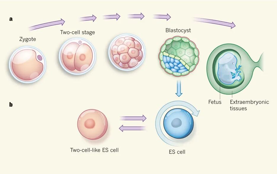
Although all cells have the same basic components, the cells in your body vary according to their specific structure and function. A zygote is the first cell formed when a sperm and an egg unite during fertilization. This first single cell will grow and then split into two cells, which will also grow and “stem off” into two cells, and so on. The process continues, forming many copies of that first cell. These cells are at first unspecialized. Unspecialized cells are called stem cells, which can differentiate (or change into) specialized cells. The image below shows a later stage of this process in which a stem cell gives rise to one cell that specializes into a neuron or other type of body cell and another that remains a stem cell.

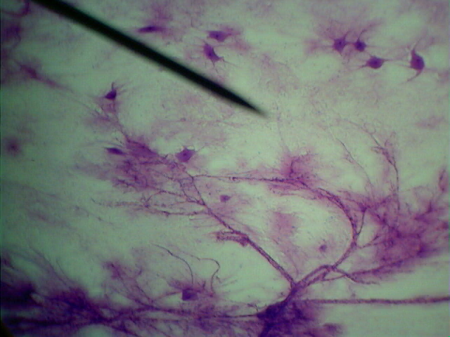
Specialized cells are different from one another in structure and function. Just like doctors specialize in different areas of medicine, such as a heart specialist, a neurosurgeon, or a family doctor, cells specialize and become experts at their specific job. For example, neurons are nerve cells that are important for all communication processes in your body and must stretch parts of themselves in many directions to send and receive signals. Neurons have many branching “limbs” called dendrites and long cytoplasm extensions called axons to transmit messages from your skin to your brain, such as the sensation of wearing soft gloves on your hands. Neurons also transmit all other sensations, including pressure, pain, heat, cold, odours, tastes, sights, and sounds. This drawing is a typical neuron.

The following table summarizes some of the different cells found in your body, and their specific function.
| Type of cell | Shape and structure | Function | Diagram/Image |
|
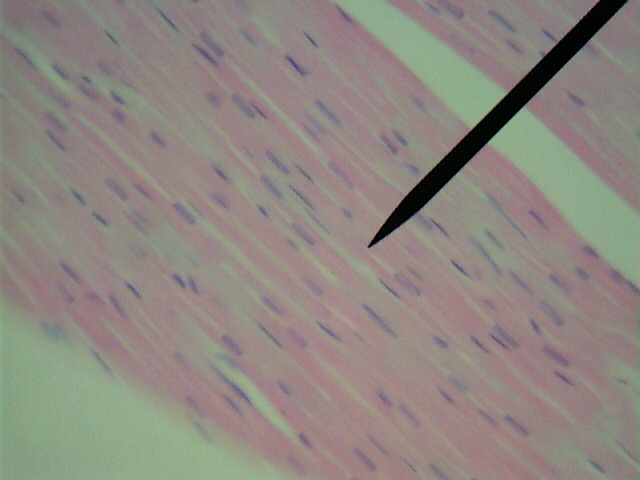
Muscle |
Elongated and often striped, muscle cells are tapered at either end. |
Contraction: Muscle cells move parts of the body by quickly contracting. Muscle cells can change shape from long and thin to short and fat. |
|
|
Skin |
Flat and thin, brick-shaped or honeycomb-shaped. Skin cells are very tough. |
Protection: Skin cells fit closely together to form several continuous protective layers. Skin layers keep all but the sharpest of objects from entering the body and introducing infection. |
|
|
Nerve |
Nerve cells have several dendrites—the long branched fibres running from the main part of the cell. |
Communication: A neuron’s dendrites carry nerve signals from one part of the body to another. In some human nerve cells, they can be over one metre long. |
|
|
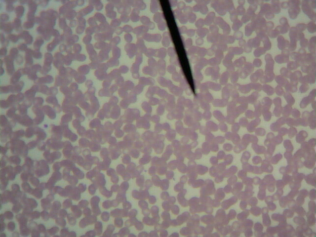
Red Blood Cells |
Blood cells are thin and disc-like, and often resemble a doughnut because light passes through the thinnest part of the cell. |
Circulation: Because blood cells must carry oxygen in the bloodstream, their flattened shape gives them a large surface area for quickly collecting oxygen, when passing near the lungs. |
|
|
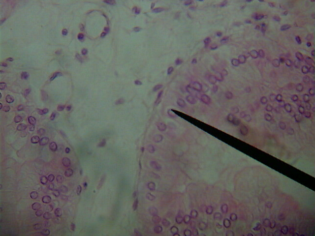
Bone |
Bone cells surround themselves with a thick mineral and protein matrix. |
The thick layer of calcium deposits secreted by osteoblasts (bone cells), provide your body with internal support. Without bones you would resemble a limp bag of jelly. This photo shows osteoclasts which are responsible for absorbing bone tissue during growth and healing. |

|
A single-celled organism is more limited than a larger, multicellular organism due to its lack of co-ordinated systems, organs, and cells. Not only are specialized cells important for specific functions within your body, but organisms with specialized cells can also:
- live in a wide variety of environments
- grow very large
- obtain their energy from a wide variety of foods
- have complex bodies
Specialized cells are dependent on other cells to survive and to perform essential functions. When you touch your hand to your face, nerve impulses originate in your brain, making your arm muscles contract. This moves your forearm bones toward your upper-arm bones, bringing your hand to your face. Often, specialized cells work together to allow the organism to function as a whole. If nerve cells in your heart stopped working, your whole body would stop working. For example, at least once a second your nerve cells send a message to the muscle cells in your heart to contract and pump blood throughout your body. If your heart stopped pumping blood, your brain cells would stop getting oxygen and you would lose consciousness.
Cancer cells are a type of specialized cell that has mutated. They stop performing their original function and start producing more cancerous cells. Unlike most of the specialized cells in your body, cancer cells do not operate in harmony with your body. This will be discussed in more detail throughout Unit 1.
Try It!
Review what you have you studied in this learning activity and answer the following questions.
Explain how the shape of a red blood cell helps with its functions carrying oxygen.
Because it is round or disc shaped, it has a large area to carry oxygen.
Identify the type of cells shown in the following image and describe its function or importance in one sentence
The image shows bone cells. Bone cells are important for giving the body support and they actively heal and allow for growth.
Reflection
Self-check
Do a self-check here. Make sure you have done, or can do, the following:
- I have set up a notebook (pen and paper or digital) to take notes and make reflections throughout the course
- I have reflected on the content in this learning activity and have reviewed key concepts
- I have begun thinking about the upcoming culminating activity and have looked ahead to learning activity 4.7 for a full description of the activity as well as the rubric that will be used to assess my work